
ELISA and other immunoassays are used to detect and measure proteins, antibodies, and other substances in biological samples. ELISA immunoassay kits have a crucial role to play for consistent and reproducible results in experiments.
Fundamentals
Immunoassays rely on a highly specific and strong interaction between antigen and antibody as a “lock and key” mechanism. An antibody has a binding region called a paratope that precisely binds to an epitope on an antigen.
Driven by high-affinity, non-covalent forces, this interaction ensures exceptional selectivity to target a molecule, keeping false signals to an absolute minimum.
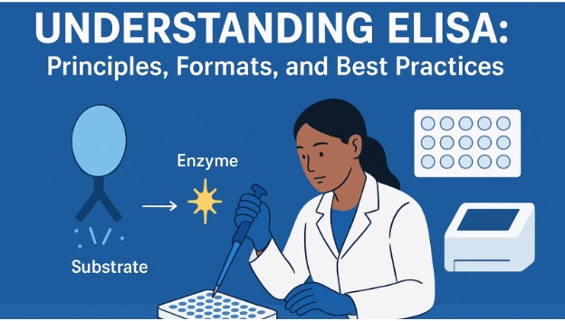
A special enzyme, such as horseradish peroxidase (HRP), attached to the antibody makes this binding visible and quantifiable. After this binding takes place, a clear liquid called substrate is introduced.
The enzyme attached to the antibody catalyzes a chemical reaction that converts the substrate into a visual signal. It can be a:
- Colorimetric Signal (substrate converted to a colored product)
- Chemiluminescent Signal (substrate converted to a product that emits light)
- Fluorescent Signal (substrate converted to a product that fluoresces when excited by a specific wavelength of light)
The signal is then measured using a microplate reader. The intensity of the signal quantifies the captured target antigen.
In the end, researchers use the standard curve to translate the raw signal into a precise measurement of concentration.
ELISA Formats
The choice of the right ELISA format depends on the target analyte and the desired outcomes in the experiment. Each format offers unique advantages.
Direct ELISA
A single enzyme-linked primary antibody directly binds to the target antigen on the plate. While the risk of cross-reactivity in this fast and simple format is low, it is less sensitive. Moreover, each target requires a unique, labeled primary antibody, which can be expensive. Direct ELISA is best for fast screening and quality control of purified antigens.
Indirect ELISA
First of all, an unlabeled primary antibody binds to an immobilized target antigen. After that, an enzyme-linked secondary antibody binds to the primary antibody. The enzyme-linked secondary antibody amplifies the signal. It is also cost-effective and versatile. However, the use of secondary antibodies can introduce the risk of non-specific binding. Indirect ELISA is ideal for serological assays and determining antibody presence and titer.
Sandwich ELISA
This format is used to detect analytes that have at least two different epitopes. It uses two antibodies:
- A capture antibody
- A detection antibody
The capture antibody is immobilized on the plate and binds to one epitope on the analyte, immobilizing it. The detection antibody, which is enzyme-linked, then binds to a different epitope on the same analyte.
The high specificity and sensitivity of this method make it ideal for quantitative analysis. However, it requires a validated pair of antibodies that recognize different epitopes. It’s an ideal method for the accurate quantification of large proteins in complex samples.
Competitive ELISA
A known amount of enzyme-linked antigen and the sample’s analyte compete for a limited binding site. It is ideal for detecting small molecules with a single binding site. The signal is inversely proportional to analyte concentration. However, it requires careful validation of competing components. It is used for the analysis of small molecules, drug detection and hormone assays.
Protocol Optimization and Best Practices
It is crucial to pay attention to protocol and adopt best practices to ensure reliable and reproducible ELISA results.
Reagent Selection & Purity
You need to minimize non-specific binding and ensure consistent performance. Make sure you use high-grade, validated reagents, pure antibodies, blocking agents, and substrates. Buy ELISA immunoassay kits and other supplies from a certified supplier.
Standard Curve Generation
You need a robust standard curve for accurate quantification. This curve represents the signal of known analyte concentrations, allowing you to determine the concentration of your unknown samples.
Advanced fitting models like the 4-parameter logistic (4-PL) generate a highly accurate curve. The R-squared value of 0.99 or higher ensures a reliable representation of the data.
Troubleshooting Common Issues
High Background
This issue occurs due to incomplete washing or ineffective blocking, a crucial step that prevents non-specific binding. Thorough washing of unbound reagents can help in avoiding high background. Use a higher concentration of blocking agent or a different blocking buffer.
Low Signal
This issue occurs due to the following reasons:
- Inefficient plate coating
- Degraded reagents
- Improper incubation times
Reagents can be sensitive to light and temperature. So, follow the manufacturer’s instructions for storage. Make sure that primary and secondary antibodies are compatible. Perform all incubation steps for the recommended duration.
Matrix Effects
Components in complex biological samples can interfere with antibody binding and lead to inaccurate results. Perform serial dilutions of the samples.